This site does not support Internet Explorer.

Try viewing this page in a different browser. All modern browsers, including Chrome, Edge, Vivaldi, Opera, Firefox, Waterfox, Tor Browser, and Safari, are supported.

Written by Eric Morshed

Published on

Cover image credit: Fayette A Reynolds/Berkshire Community College. Public domain.

Before inventing the most popular system used by blind people to read, Louis Braille was a young, well‐sighted lad who loved taking after his father.

His father spent a lot of his time working in his shed. He made saddles, stirrups, and all sorts of other things that needed some fancy tools. The young Louis Braille was very fascinated by these tools, tried to use them like his father did and it was all fun and games until he hit himself in the eye.

It was not a very pretty scene, nor a particularly fun experience for Braille himself. The damage to his right eye proved unsalvageable, but at least he had his other eye to see with — until something unfortunate happened to it, as well.

The eyes have a direct wire to the brain and the central nervous system (CNS), and like the rest of the CNS they are afforded some special privileges by the immune system. Unfortunately, an injury to the eye puts an end to this privilege: if the physical barriers that keep the most aggressive parts of the immune system out of the eye are breached, a condition called sympathetic ophthalmia can occur. The immune system, seeing the interior environment of the eye for the first time, gets confused and attacks it, wreaking havoc on vision. This, it is believed, is the unfortunate fate that befell Louis Braille’s other eye.1

Ever since the discovery of this separation between the immune system and the CNS — the blood–brain and blood–eye barriers (BBB and BEB, respectively), for instance — the prevailing thought has been that the immune system and the nervous system are all but anathema to each other, kept completely and intentionally separate for the brain’s own safety. In the past decade or so, however, mounting evidence has begun to show that this is not at all the case. The relationship between the brain and immunity is much more nuanced, and in it, perhaps, lie the secrets behind countless mysterious diseases.

1 A Cloak of Invi­si­bility

The “old view.” so to speak, is not without its merits. Many components of the immune system simply don’t operate in the brain at the same capacity as they do in the rest of the body, and there are plenty of good reasons for this. For instance, although new neurons can be produced over the course of a person’s lifetime, this generally happens at an incredibly slow pace,2 and so any nerve cells that are destroyed as collateral damage in an immune response — very much a possibility, considering that the immune system sometimes likes to carpet‐bomb the body to get rid of threats — would be difficult to replace.

Instead, to protect itself from disease, the nervous system relies mainly on physical barriers like the aforementioned BBB, which consists of cells like astrocytes and pericytes that surround blood vessels in the brain and have extremely narrow gaps between them. The idea is that the brain doesn’t need to bother with massive immune responses if germs are too fat to squeeze through the gaps in the BBB and enter the brain in the first place.

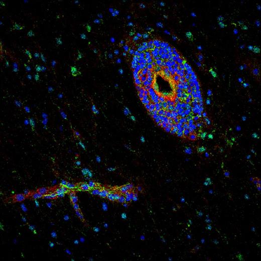
Layers of fluorescent blue and green cells surrounding a hollow blood vessel

This micrograph shows the layers of cells coating blood vessels that comprise the BBB.

Credit: Podjaski et al., 2015.

Generally, the BBB does quite a good job, keeping out not only pathogens but all kinds of other things — acting, if you will, like an invisibility cloak for the brain. For instance, the BBB was first noticed by scientists who found that dyes injected into the blood would seemingly touch every part of the body but the brain.3 And we have pretty solid indications that the brain relies on the BBB doing its job. Factors such as aging and exposure to chemical weapons can lead to a permanent deterioration of the BBB, allowing inflammatory responses from the immune system to intrude upon the brain and bring about cognitive decline.4; 5 Such BBB dysfunction is even believed to be a significant contributor to cognitive decline in Alzheimer’s disease. The exact reasons for this are under investigation and up for interpretation, but it at least partially has to do with inflammatory responses that enter the brain as the BBB falls apart.5; 6 The fact that this doesn’t normally happen is a testament to the BBB’s importance and how well it does its job in healthy individuals.

As foolproof as the BBB’s approach may seem, it has one insidious weakness. By necessity, the brain is connected to the rest of the body by nerves. Also by necessity, many of these nerves are not as well‐shielded from disease as the brain itself. A particularly determined pathogen could, therefore, enter a nerve and gradually work its way up into the brain.

The most infamous examples of this are lyssaviruses, such as the one that causes rabies. Upon entering a nerve, they crawl along the intracellular transport networks of the neurons there, hopping slowly but determinedly from cell to cell until they reach the brain7. A similar approach can be used by herpes simplex virus, resulting in encephalitis.5; 8

Furthermore, the brain can face threats from within. Normal cellular functions produce massive amounts of waste that can prove toxic if allowed to accumulate, and that therefore need to be cleaned up somehow. Just like any other part of the body, the brain is also subject to physical injury, which can cause cell damage that needs to be cleaned up. And, of course, there can always be more extreme conditions like rogue cancerous cells. When problems like these occur, the BBB is powerless to do anything — at the end of the day, it is just a passive barrier. The brain needs an active helper, and so the various components of the immune system must enter the brain to take care of disease, waste disposal, and other tasks.

So how do they get there? As is typical for seemingly simple questions, the answer to that question is … it depends.

2 Ladon

How the immune system enters the brain depends on a lot of different factors: the type of immune cell, the circumstances the body is under, and more. In one specific case, though, the answer is quite simple: they don’t get there. Some of them are simply always there.

Destined to be born and die in the brain are a determined contingent of special immune cells known as microglia. They — or at least their ancestors, such as yolk sac myeloid progenitor cells — arrive in the brain extremely early in embryonic development and live out the rest of their lives there.9 They take care of everyday functions like removing waste and even playing an important role in learning by pruning off the dead weight of unnecessary connections between neurons. Often, they are rather benign in their operations, keeping collateral damage to a necessary minimum.

That is except when they aren’t.

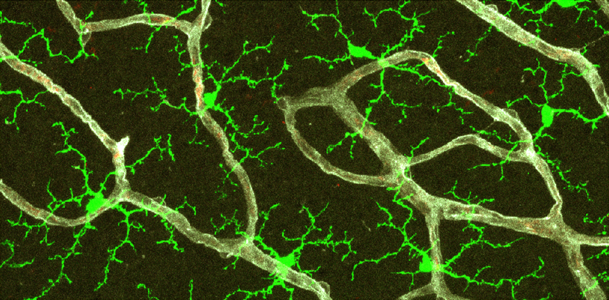
Neon green cells with branching networks emerging from them

The heroes of the upcoming story: microglia, stained here in bright green.

Credit: Wenshin Ma and Wai Wong/National Eye Institute. Public domain.

Having toothless immune cells is rather pointless — microglia can bare their teeth when needed. In the brain and in the neural tissue of the eyes, they have the ability to promote inflammatory responses as well as to kill cells when necessary.10 At the same time, microglia have the capacity to encourage the development of new neurons to replace ones that may be killed by injury or by immune responses.11 The balance between these creative and destructive forces is one that must be carefully maintained. When the balance fails to work out, unabated destruction of neurons can ensue. In the brain, this can result in cognitive decline; in the eyes, the consequence is macular degeneration, permanently impairing vision.10 Maintaining this balance is where microglia’s friends enter the picture.

3 Tra­vel­ers from Afar

Though microglia are the native immune cells of the nervous system, recent findings show that they are often not alone. In the healthy brain, a very limited and carefully‐controlled cadre of immune cells from the rest of the body are allowed to sneak through the BBB at the behest of microglia, primarily comprising macrophages, monocytes, and Th cells.12

Macrophages and monocytes — they are quite similar — are some of the most common immune cells in the body, and upon being allowed to enter the brain they carry out several critical functions. Like microglia, most of their work is swallowing up waste and debris to be digested. Their work is so similar to that of microglia, in fact, that it appears that some of them actually stay in the brain, specializing to become microglia themselves.9

The other critical role these macrophages have, and one they share with the Th cells, is surveillance. These immune cells belong to a category of cells known as professional antigen presenting cells — yes, that is the actual technical name. The “professional” part of the name comes from the fact that they have one very specific job: picking up antigens, which are molecular keepsakes they find from their environment, and “presenting” them to other immune cells that can identify whether or not those antigens are signs of a threat. To that end, these friends of microglia creep slowly in and out of the brain, surveilling it for omens of trouble.12

Macrophages and monocytes — they are quite similar — are some of the most common immune cells in the body,13 and upon being allowed to enter the brain they carry out several critical functions. Like microglia, most of their work is swallowing up waste and debris to be digested. Their work is so similar to that of microglia, in fact, that it appears that some of them actually stay in the brain, specializing to become microglia themselves.9

In either case — whether by immune cells entering the brain directly or by samples of brain juices being smuggled out by glymphatic flow for immune inspection — a small army of immune cells waits for the opportunity to raise the alarm when a threat arises. When they do, everything changes.

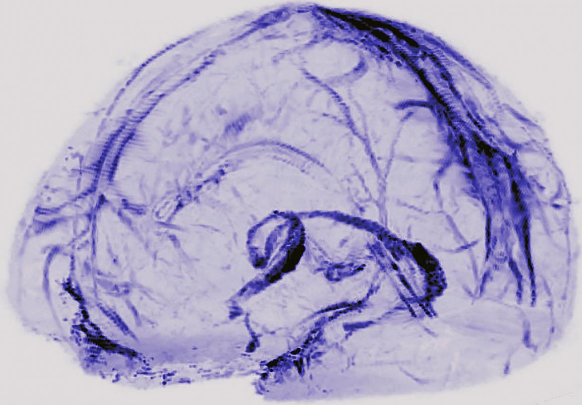
Blue branching structures outlining the shape of a brain

In collaboration with the glymphatic system, these recently discovered lymph vessels right outside the brain allow for immune surveillance of what goes on inside the BBB.

Credit: Reich Lab, National Institute of Neurological Disorders and Stroke. Public domain.

The signals released by panicked microglia cause changes in the BBB that make it far more conducive to being crossed by immune cells from all across the body, including ones that would otherwise stay out of the nervous system entirely. One of the most remarkable guests invited are B cells,12 which produce antibodies: proteins that recognize antigens and flag them for attention from the immune system. Though they form a necessary part of some immune responses, the wrath of B cells can also be harmful when turned against the brain itself. In fact, such B cell infiltrations of the nervous system are implicated in notorious diseases like multiple sclerosis,14 since they expose the brain to the most direct form of assault the immune system has to offer.

Throughout all of this, one important concept rings clear: balance. All these ventures of the immune system into the brain are, at times, necessary — but their destructive potential can wreak havoc on important bodily functions. In itself, that is not too out of the ordinary. There is an entire category of so‐called autoimmune diseases that revolve around the immune system attacking the body. Even in non‐autoimmune diseases like COVID‐19, an overly aggressive immune response can sometimes hurt the body more than the disease itself.15 An imbalance in the other direction can be equally deadly — many cancers, for instance, eke out a living by convincing the immune system to leave them alone.15 When they occur elsewhere in the body, these imbalances can be treated with various kinds of drugs, from immunosuppressants used to calm down the immune system to artificial antibodies used to reinvigorate it. The trouble with the CNS, though, is that when the immune system goes awry, the very thing that is supposed to keep it under control — the BBB — gets in the way by blocking such drugs from entering the brain. Workarounds are under investigation, but they are fundamentally still difficult.17 Even with these workarounds, it often appears that trying to beat the immune system into submission with such conventional drugs just straight‐up doesn’t work in the brain, for reasons that are not entirely clear.18

So perhaps, instead of working against the unique properties of the neurological immune system, we must learn to work with it.

4 Takes One to Know One

Immunotherapy has been picking up momentum for the last decade or so, and recently, attempts have been made to extend it into the unique environment of the nervous system. Primarily, these efforts have focused on the star of the show: microglia.

“Reprogramming” a patient’s immune cells made its debut into the mainstream medical scene with CAR‐T cell therapy, often used to fight cancer. Such a therapy entails extracting a sample of a patient’s T cells and genetically engineering them with chimeric antigen receptors (CARs) that can recognize cancer antigens, after which the CAR‐T cells can be reinjected into the body to go serve justice. The same type of approach can be used with microglia. Techniques to either edit microglial genes or change how often they are used, or expressed, in the technical parlance, can convert these cells between the immune‐activating type M1 and the immune‐suppressing type M2, thereby providing a way to restore balance in the case of diseases where it has been lost.19; 20 Various different methods can be used for this purpose. To promote M2 microglia, we can engineer microglia to simply not have the genes necessary for inflammation, or we can engineer macrophages that attack and disable pro‐inflammatory microglia. Conversely, M1 microglia can be promoted by doing just about the opposite: genetically engineering them to promote inflammation where they otherwise wouldn’t, for instance.20

Nevertheless, the same fundamental problem remains of getting things through the BBB. Injecting genetically‐engineered cells directly into the nervous system is risky, to say the least. Fortunately, though, this might not be necessary. Recall that when inflammation is already underway, immune cells from outside the CNS are able to enter it even when other substances are still kept out. To reduce inflammation, therefore, it would suffice to inject anti‐inflammatory cells anywhere in the body, whereupon they can find their way to the appropriate location — aided, perhaps, by genetic engineering — and cross the BBB to put an end to inflammation in the brain. This approach has been tried in mice and other animals using various kinds of anti‐inflammatory cells, such as macrophages21 and — uniquely — Treg (regulatory T) cells, which exist for the sole purpose of downregulating immune responses. As their similar name would suggest, Treg and Th cells are quite similar, and they can presumably enter the brain through similar mechanisms. Once there, they faithfully perform their duty of suppressing immune responses, both deterring immune cells that enter the brain from the outside and encouraging microglia to convert to the M2 type.22 Research into these therapies is still underway, but on the whole, it seems that taking advantage of the BBB’s willingness to let immune cells into the inflamed brain allows for remarkably effective therapy to reduce inflammation.

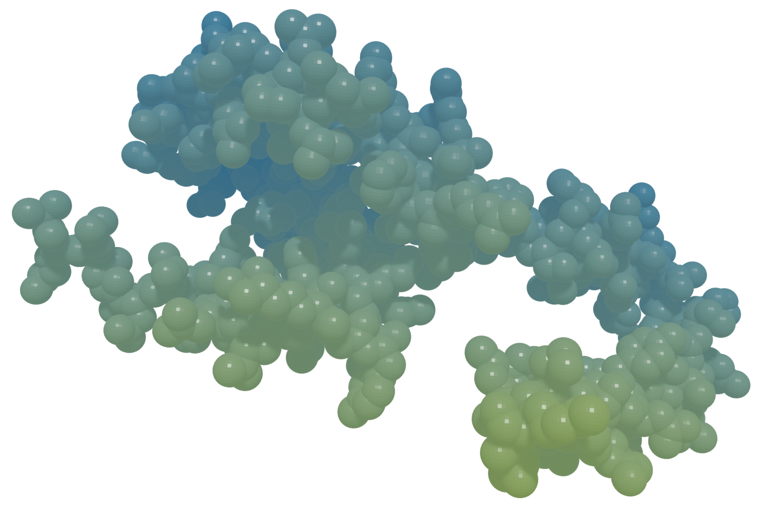
3D render of protein structure

Interleukin‐10, one of the major immunoregulatory proteins produced by Treg cells.

Credit: Original graphic, based on Protein Data Bank entry 2H24.

The opposite task, meanwhile, seems comparatively more difficult. Not only is it hard to sneak things past the BBB when inflammation is low, but promoting inflammation can also be risky if it is done indiscriminately due to the possibility of damaging important neural tissue. As such, promoting immune responses must be done in a very careful and specific way.

Th cells provide a promising way of doing this. Consider CAR‐T cell therapy, for example. Adapting this therapy to brain cancers like glioblastoma has been difficult due to the intricacies of the neuroimmune system, but recently, two independent teams of researchers finally cracked the code — at least in mice.23; 24 In both cases, the CAR‐T cells were administered directly into the fluid surrounding the brain and spinal cord, bypassing the BBB altogether, but there is no reason to believe that this is the only way. We have seen artificially‐administered T cells entering the brain in experimental autoimmune encephalomyelitis, which is a sort of “simulation” of multiple sclerosis in animals like mice.(25) To further encourage CAR‐T cell entry into the CNS, the cells could be genetically engineered to express greater levels of the immune cell transport proteins like laminin‐411 and CD166 that are specifically relevant when crossing the BBB.12

Ideas like the ones we’ve discussed lie on the bleeding edge of medicine. So much of what we’ve covered here has been only very recently discovered, upending the tacit assumption that, between the CNS and the full gamut of the immune system, never the twain shall ever meet.

But already, we are seeing the immense potential of medically assisting the careful balancing act of the neuroimmune system. In the secret codewords passed by and between cells across the no‐man’s‐land that is the BBB, there is a chance to sneak some messages to the neuroimmune system ourselves. To halt its self‐destructive tendencies, or to awaken it when it slumbers. To give it guidance to fight everything from injuries to horrifying neurodegenerative diseases to age‐related cognitive decline. What was once a medical unknown could become one of our greatest allies.


D Dedi­cation

This article is dedicated to Santiago Ramón y Cajal (1852–1934), whose work on the cellular anatomy of the central nervous system arguably founded the modern field of neuroscience. Early on in his life, his destiny was far from predetermined: he was born into no great wealth in the Spanish town of Petilla de Aragón, apprenticed with cobblers and barbers, and wanted to be an artist. At the behest of his father, the surgeon of their village, he was made to study medicine at the University of Zaragoza. There, he set upon the idea of applying his artistic talents to the mysteries of biology.26

In 1887, an opportunity presented itself. Ramón y Cajal was visiting Luis Simarro Lacabra, a neurologist working out of Madrid. There, he saw something marvelous: a particular method of treating nervous tissue, a slight modification made by Simarro to an innovative method invented a few years prior by the Italian biologist Camillo Golgi. This method allowed for the cells within the tissue to be visualized under a microscope with astonishing and previously unachievable clarity: for the first time, it was possible to actually investigate how the brain works on a cellular level.27; 28 Ramón y Cajal immediately began using this approach in his own work, creating beautiful and scientifically rigorous depictions of neurons and making earth‐shattering discoveries that are taken for granted today. For instance, he was the first to point out that neurons do not merge and meld into each other, as Golgi thought, but rather are independent cells, capable of dynamically forging connections to others.27; 29 He also made the key realization that studying the brains of embryos would be greatly insightful, since, in these early stages of development, the complex webs of connections between neurons are, well, less complex and easier to understand.29 For this work, he shared the 1906 Nobel Prize for Physiology or Medicine with Golgi.26

Ramón y Cajal’s legacy has proved long‐lasting. Not only was his own work foundational to modern neuroscience, but his eagerness to share his knowledge spawned even more innovations. In fact, it was one of his students — Pío del Río‐Hortega — who characterized microglia and discovered their role as the immune cells of the brain,30 kicking off the curious tale of the neuroimmune system.

S Sources

  1. “Louis Braille: Dancing in the Dark” by Mrittika Sen, Santosh G Honavar. Vol. 70, no. 1, pp. 5–6 from: Indian Journal of Ophthalmology. Published on behalf of the All Indian Ophthalmological Society (Delhi, DL, IN) by Wolters Kluwer (Alphen aan den Rijn, ZH, NL) in 2022 Jan. URL: https://journals.lww.com/ijo/Fulltext/2022/01000/Louis_Braille__Dancing_in_the_Dark.3.aspx; DOI: 10.4103/ijo.IJO_3048_21; PMID: 34937200; PMCID: PMC8917520.

  2. “Generation of Neurons in the Adult Brain.” Ch. 25 from: Neuroscience (2nd ed.) by Dale Purves, George J Augustine, David Fitzpatrick, Lawrence C Katz, Anthony‐Samuel LaMantia, et al. Published by Sinauer Associates (Sunderland, MA, US) in 2001. URL: https://www.ncbi.nlm.nih.gov/books/NBK10920/; ISBN: 0-87893-742-0.

  3. “History of Blood–Brain Barrier.” from: The Arizona Brain Barriers Laboratory. Published by the University of Arizona (Tucson, AZ, US); copyright 2021; accessed 2024 Jul 19. URL: https://davislab.med.arizona.edu/history-blood-brain-barrier.

  4. “Damage to a Protective Shield around the Brain May Lead to Alzheimer’s and Other Diseases” by Daniela Kaufer, Alon Friedman. Vol. 325, no. 5 from: Scientific American. Published by Springer Nature (London, ENG, UK) on 2021 May 01. URL: https://www.scientificamerican.com/article/damage-to-a-protective-shield-around-the-brain-may-lead-to-alzheimers-and-other-diseases/; DOI: 10.1038/scientificamerican0521-42.

  5. “Blood–Brain Barrier Breakdown: An Emerging Biomarker of Cognitive Impairment in Normal Aging and Dementia” by Basharat Hussain, Cheng Fang, Junlei Chang. Vol. 15, art. 688090 from: Frontiers in Neuroscience. Published by Frontiers Media (Lausanne, VD, CH) on 2021 Aug 18. URL: https://www.frontiersin.org/journals/neuroscience/articles/10.3389/fnins.2021.688090/full; DOI: 10.3389/fnins.2021.688090; PMID: 34489623; PMCID: PMC8418300.

  6. “APOE4 Leads to Blood–Brain Barrier Dysfunction Predicting Cognitive Decline” by Axel Montagne, Daniel A Nation, Abhay P Sagare, Giuseppe Barisano, Melanie D Sweeney, et al. Vol 581, pp. 71–76 from: Nature. Published by Springer Nature (London, ENG, UK) on 2020 Apr 29. URL: https:////pmc.ncbi.nlm.nih.gov/articles/PMC7250000/; DOI: 10.1038/s41586-020-2247-3; PMID: 32376954; PMCID: PMC7250000.

  7. “Interaction of the Rabies Virus P Protein with the LC8 Dynein Light Chain” by Hélène Raux, Anne Flamand, Danielle Blondel. Vol. 74, no. 21 from: Journal of Virology. Published by the American Society for Microbiology (Washington, DC, US) on 2000 Nov 01. URL: https://journals.asm.org/doi/10.1128/jvi.74.21.10212-10216.2000; DOI: 10.1128/jvi.74.21.10212-10216.2000; PMID: 11024151; PMCID: PMC102061.

  8. “Portals of Viral Entry into the Central Nervous System” by Phillip A Swanson II, Dorian B McGavern. Vol. 2, ch. 2 from: The Blood–Brain Barrier in Health and Disease, edited by Katerina Dorovini‐Zis. Published by the CRC Press (Boca Raton, FL, US) on 2015 Oct 06. URL: https://www.researchgate.net/publication/282851330_Portals_of_Viral_Entry_into_the_Central_Nervous_System; DOI: 10.1201/b19299; ISBN: 978-0-42-907579-7.

  9. “Origin and Emergence of Microglia in the CNS — An Interesting (Hi)story of an Eccentric Cell” by Iasonas Dermitzakis, Maria Eleni Manthou, Soultana Meditskou, Marie‐Ève Tremblay, Steven Petratos, et al. Vol. 45, no. 3, pp. 2609–2628 from: Current Issues in Molecular Biology. Published by MDPI (Basel, BS, CH) on 2023 Mar 22. URL: https://www.mdpi.com/1467-3045/45/3/171; DOI: 10.3390/cimb45030171; PMID: 36975541; PMCID: PMC10047736.

  10. “The TSPO–NOX1 Axis Controls Phagocyte‐Triggered Pathological Angiogenesis in the Eye” by Anne Wolf, Marc Herb, Michael Schramm, Thomas Langmann. Vol. 11, art. 2709 from: Nature Communications. Published by Springer Nature (London, ENG, UK) on 2020 Jun 01. URL: https://www.nature.com/articles/s41467-020-16400-8; DOI: 10.1038/s41467-020-16400-8; PMID: 32483169; PMCID: PMC7264151.

  11. “Microglia Activated by IL‐4 or IFN‐γ Differentially Induce Neurogenesis and Oligodendrogenesis from Adult Stem/Progenitor Cells” by Oleg Butovsky, Yaniv Ziv, Adi Schwartz, Gennady Landa, Adolfo E Talpalar, et al. Vol. 31, no. 1, pp. 149–160 from: Molecular and Cellular Neuroscience. Published by Elsevier (Amsterdam, NH, NL) in 2006 Jan. URL: https://www.sciencedirect.com/science/article/abs/pii/S1044743105002484; DOI: 10.1016/j.mcn.2005.10.006; PMID: 16297637.

  12. “Immune Cell Trafficking across the Blood–Brain Barrier in the Absence and Presence of Neuroinflammation” by Luca Marchetti, Britta Engelhardt. Vol. 2, no. 1, pp. H1–H18 from: Vascular Biology. Published by Bioscientifica (Bristol, ENG, UK) on 2020 Apr 10. URL: https://vb.bioscientifica.com/configurable/content/journals$002fvb$002f2$002f1$002fVB-19-0033.xml; DOI: 10.1530/VB-19-0033; PMID: 32923970; PMCID: PMC7439848.

  13. “The Glymphatic System” by Lauren M Hablitz, Maiken Nedergaard. Vol. 31, no. 20, pp. R1371–R1375 from: Current Biology. Published by Cell Press (Cambridge, MA, US) on 2021 Oct 25. URL: https://www.cell.com/current-biology/fulltext/S0960-9822(21)01130-1; DOI: 10.1016/j.cub.2021.08.026; PMID: 34699796.

  14. “Activated Leukocyte Cell Adhesion Molecule Regulates B Lymphocyte Migration across Central Nervous System Barriers” by Laure Michel, Camille Grasmuck, Marc Charabati, Marc‐André Lécuyer, Stephanie Zandee, et al. Vol. 11, no. 518 from: Science Translational Medicine. Published by the American Association for the Advancement of Science (Washington, DC, US) on 2019 Nov 13. URL: https://www.science.org/doi/10.1126/scitranslmed.aaw0475; DOI: 10.1126/scitranslmed.aaw0475; PMID: 31723036.

  15. “Cytokine Storm” by David C Fajgenbaum, Carl H June. Vol 383, no. 23 from: The New England Journal of Medicine. Published by the Massachusetts Medical Society (Waltham, MA, US) on 2020 Dec 02. URL: https://www.nejm.org/doi/full/10.1056/NEJMra2026131; DOI: 10.1056/NEJMra2026131; PMID: 33264547; PMCID: PMC7727315.

  16. “How Cancers Escape Immune Destruction and Mechanisms of Action for the New Significantly Active Immune Therapies: Helping Nonimmunologists Decipher Recent Advances” by Jonathan L Messerschmidt, George C Prendergast, Gerald L Messerschmidt. Vol. 21, no. 2, pp. 233–243 from: The Oncologist. Published by the Oxford University Press (Oxford, ENG, UK) on 2016 Feb 01. URL: https://academic.oup.com/oncolo/article/21/2/233/6401118; DOI: 10.1634/theoncologist.2015-0282; PMID: 26834161; PMCID: PMC4746082.

  17. “Chemotherapy and Immunosuppressant Therapy–Induced Posterior Reversible Encephalopathy Syndrome” by Gurleen Kaur, Ibtisam Ashraf, Mercedes Maria Peck, Ruchira Maram, Alaa Mohamed. Vol. 12, no. 10, art. e11163 from: Cureus. Published on behalf of the California Institute of Behavioral Neurosciences & Psychology (Fairfield, CA, US) by Springer Nature (London, ENG, UK) on 2020 Oct 25. URL: https://www.cureus.com/articles/40643-chemotherapy-and-immunosuppressant-therapy-induced-posterior-reversible-encephalopathy-syndrome; DOI: 10.7759/cureus.11163; PMID: 33251070; PMCID: PMC7688184.

  18. “M1/M2 Polarization in Major Depressive Disorder: Disentangling State from Trait Effects in an Individualized Cell‐Culture–Based Approach” by Nicoleta Carmen Cosma, Berk Üsekes, Lisa Rebecca Otto, Susanna Gerike, Isabella Heuser, et al. Vol 94, pp. 185–195 from: Brain, Behavior, and Immunity. Published by Elsevier (Amsterdam, NH, NL) in 2021 May. URL: https://www.sciencedirect.com/science/article/pii/S0889159121000519; DOI: 10.1016/j.bbi.2021.02.009; PMID: 33607231.

  19. “Targeting Neuro–Immune Communication in Neurodegeneration: Challenges and Opportunities” by Aleksandra Deczkowska, Michal Schwartz. Vol. 215, no. 11, pp. 2702–2704 from: Journal of Experimental Medicine. Published by the Rockefeller University Press (New York, NY, US) on 2018 Oct 09. URL: https://rupress.org/jem/article/215/11/2702/120279/Targeting-neuro-immune-communication-in; DOI: 10.1084/jem.20181737; PMID: 30301785; PMCID: PMC6219738.

  20. “Taming Microglia: The Promise of Engineered Microglia in Treating Neurological Diseases” by Echo Yongqi Luo, Rio Ryohichi Sugimura. Vol. 21, art. 19 from: Journal of Neuroinflammation. Published by BioMed Central (London, ENG, UK) on 2024 Jan 11. URL: https://jneuroinflammation.biomedcentral.com/articles/10.1186/s12974-024-03015-9; DOI: 10.1186/s12974-024-03015-9; PMID: 38212785; PMCID: PMC10785527.

  21. “Photoresponsive Vaccine‐Like CAR‐M System with High-‐Efficiency Central Immune Regulation for Inflammation-Related Depression” by Yu Liu, Ping Hu, Zhiheng Zheng, Da Zhong, Weichang Xie, et al. Vol. 34, no. 11 from: Advanced Materials. Published by Wiley‐VCH (Weinheim, BW, DE) on 2021 Dec 12. URL: https://onlinelibrary.wiley.com/doi/10.1002/adma.202108525; DOI: 10.1002/adma.202108525; PMID: 34897839.

  22. “Harnessing Regulatory T Cell Neuroprotective Activities for Treatment of Neurodegenerative Disorders” by Jatin Machhi, Bhavesh D. Kevadiya, Ijaz Khan Muhammad, Jonathan Herskovitz, Katherine E Olson, et al. Vol. 15, art. 32 from: Molecular Neurodegeneration. URL: https://molecularneurodegeneration.biomedcentral.com/articles/10.1186/s13024-020-00375-7; DOI: 10.1186/s13024-020-00375-7; PMID: 32503641; PMCID: PMC7275301.

  23. “Intrathecal Bivalent CAR T Cells Targeting EGFR and IL13Rα2 in Recurrent Glioblastoma: Phase 1 Trial Interim Results” by Stephen J Bagley, Meghan Logun, Joseph A Fraietta, Xin Wang, Arati S Desai, et al. Vol. 30, pp. 1320–1329 from: Nature Medicine. Published by Springer Nature (London, ENG, UK) on 2024 Mar 13. URL: https://www.cceb.upenn.edu/ifi/assets/user-content/documents/s41591-024-02893-z.pdf; DOI: 10.1038/s41591-024-02893-z; PMID: 38480922.

  24. “Intraventricular CARv3-TEAM-E T Cells in Recurrent Glioblastoma” by Bryan D Choi, Elizabeth R Gerstner, Matthew J Frigault, Mark B Leick, Christopher W Mount, et al. Vol. 390, no. 14 from: The New England Journal of Medicine. Published by the Massachusetts Medical Society (Waltham, MA, US) on 2024 Mar 13. URL: https://www.nejm.org/doi/10.1056/NEJMoa2314390; DOI: 10.1056/NEJMoa2314390; PMID: 38477966; PMCID: PMC11162836.

  25. “The Rapid Isolation of Clonable Antigen‐Specific T Lymphocyte Lines Capable of Mediating Autoimmune Encephalomyelitis” by Avraham Ben‐Nun, Hartmut Wekerle, Irun R Cohen. Vol. 11, no. 3, pp. 195–199 from: The European Journal of Immunology. Published on behalf of the European Federation of Immunological Societies (EU) by Wiley‐VCH (Weinheim, BW, DE) in 1981 Nov. URL: https://journals.aai.org/jimmunol/article-pdf/198/9/3384/1432026/1700351.pdf; DOI: 10.1002/eji.1830110307; PMID: 6165588.

  26. “Santiago Ramón y Cajal: Biographical.” Published by Nobel Prize Outreach (Stockholm, AB, SE); accessed 2024 Jul 20. Originally published by Elsevier (Amsterdam, NH, NL) in 1967. URL: https://www.nobelprize.org/prizes/medicine/1906/cajal/biographical/.

  27. “Golgi and Cajal: The Neuron Doctrine and the 100th Anniversary of the 1906 Nobel Prize” by Mitch Glickstein. Vol. 15, no. 5, pp. R147–R151 from: Current Biology. Published by Cell Press (Cambridge, MA, US) on 2006 Mar 07. URL: https://www.cell.com/current-biology/fulltext/S0960-9822(06)01203-6; DOI: 10.1016/j.cub.2006.02.053; PMID: 16527727.

  28. “Luis Simarro Lacabra” by Helio Carpintero Capell. from: El diccionario biográfico español edited by Carmen Iglesias. Published by the Royal Academy of History (Madrid, MD, ES); copyright 2018; accessed 2024 Jul 20. URL: https://dbe.rah.es/biografias/14994/luis-simarro-lacabra.

  29. “The Father of Modern Neuroscience Discovered the Basic Unit of the Nervous System” by Benjamin Ehrlich. Vol. 326, no. 4 from: Scientific American. Published by Springer Nature (London, ENG, UK) on 2022 Apr 01. URL: https://www.scientificamerican.com/article/the-father-of-modern-neuroscience-discovered-the-basic-unit-of-the-nervous-system/; DOI: 10.1038/scientificamerican0422-50.

  30. “Pío del Río Hortega and the Discovery of the Oligodendrocytes” by Fernando Pérez‐Cerdá, María Victoria Sánchez‐Gómez, Carlos Matute. Vol. 9, art. 92 from: Frontiers in Neuroanatomy. Published by Frontiers Media (Lausanne, VD, CH) on 2015 Jul 06. URL: https://www.frontiersin.org/journals/neuroanatomy/articles/10.3389/fnana.2015.00092/full; DOI: 10.3389/fnana.2015.00092; PMID: 26217196; PMCID: PMC4493393.
X Dis­claimer

I am not a scientist or professional in any field. The content of this article merely expresses my personal views, opinions, and visions for the future. This content is not intended for use as professional advice on any matter.